
Sex and brains
Vive la différence!
A new technique has drawn wiring diagrams of the brains of the two sexes. The contrast between them is illuminating.

MEN and women do not think in the same ways. Few would disagree with that. And science has quantified some of those differences. Men, it is pretty well established, have better motor and spatial abilities than women, and more monomaniacal patterns of thought. Women have better memories, are more socially adept, and are better at dealing with several things at once.
There is a lot of overlap, obviously. But on average these observations are true.
Suggesting why they are true in evolutionary terms is a game anyone can play. One obvious idea is that because, in the days of hunting and gathering, men spent more time wandering away from camp, their brains needed to be adapted to able to find their way around. They also spent more time tracking, fighting and killing things, be they animals or intrusive neighbours. Women by contrast, politicked among themselves and brought up the children, so they needed to be adapted to enable them to manipulate each other’s and their children’s emotions to succeed in their world.
Finding out why sex differences are true in neurological terms—in other words, how the brain is wired up to create them—is another matter altogether. To play this game you have to have a lot of expensive kit, not just a comfortable chair from which to pontificate. And that is exactly what Ragini Verma of the University of Pennsylvania and her colleagues do have. As a result, as they outline in the Proceedings of the National Academy of Sciences, they have been able to map out differences in the ways male and female brains are cabled and match them, at least to their own satisfaction, to the stereotypes beloved of both folklore and psychology.
Sugar and spice or puppy-dogs’ tails?
Neurology has been revolutionised over the past couple of decades by a range of techniques that can scan living brains. Dr Verma’s technique of choice is diffusion tensor imaging. This follows water molecules around the brain. Because the fibres that connect nerve cells have fatty sheaths, the water in them can diffuse only along a fibre, not through the sheath. So, diffusion tensor imaging is able to detect bundles of such fibres, and see where they are going.
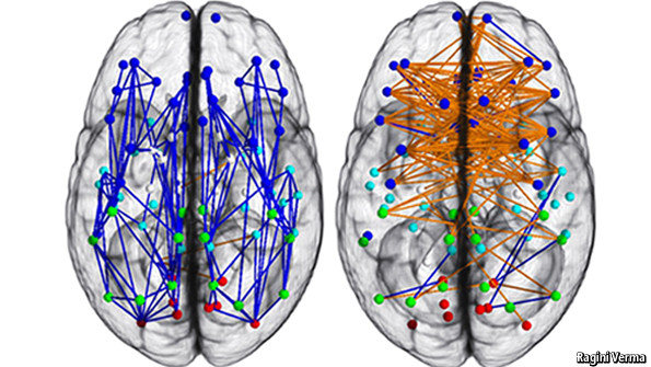
Dr Verma and her team applied the technique to 428 men and boys, and 521 women and girls. Their results are summarised in the two diagrams above, which show connection trends averaged from the sum of participants’ brains.
The two main parts of a human brain are the cerebrum, above and towards the front, which does the thinking, and the cerebellum, below and towards the back, which does the acting. Each is divided into right and left hemispheres. As the diagrams show, in men (the left-hand picture) the dominant connections in the cerebrum are those, marked in blue, within hemispheres. In women, they are those, marked in orange, between hemispheres. In the cerebellum (not visible because it is under the cerebrum), it is the other way around.
What this means is open to interpretation, but Dr Verma’s take is that the wiring differences underlie some of the variations in male and female cognitive skills. The left and right sides of the cerebrum, in particular, are believed to be specialised for logical and intuitive thought respectively. In her view, the cross-talk between them in women, suggested by the wiring diagrams, helps explain their better memories, social adeptness and ability to multitask, all of which benefit from the hemispheres collaborating. In men, by contrast, within-hemisphere links let them focus on things that do not need complex inputs from both hemispheres. Hence the monomania.
When it comes to the cerebellum, the extra cross-links between hemispheres in men serve to co-ordinate the activity of the whole sub-organ. That is important because each half controls, by itself, only one half of the body. Hence men have better motor abilities—or, in layman’s terms, are better co-ordinated than women.
Dr Verma’s other main finding is that most of these differences are not congenital. Rather, they develop with age. Her volunteers ranged from 8 to 22 years old. The brains of boys and girls aged 8 to 13 demonstrated only a few differences, though all were of the type that later became pronounced. Adolescents, those aged 13 to 17, showed more. Young adults, over 17, more still. Sex differences in brains—those visible to this technique, at least—thus manifest themselves mainly when sex itself begins to matter.
Dr Verma’s work is important not only for what it has shown, but also as a demonstration of the power of diffusion tensor imaging. Studying the brain, particularly the living brain, is a uniquely hard scientific problem. The American government has recently promised to spend serious amounts of money doing so, through what it dubs the BRAIN initiative—the inevitable contrived acronym supposedly standing for Brain Research through Advancing Innovative Neurotechnologies.
As that name suggests, advances in brain research depend on the development of better ways of looking at brains while they are alive and firing. This will be hard. But work like Dr Verma’s shows the rewards of doing it.
No comments:
Post a Comment